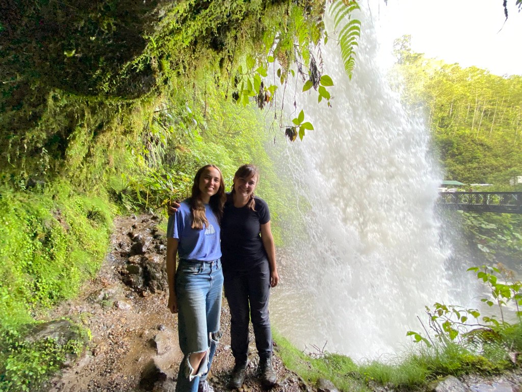
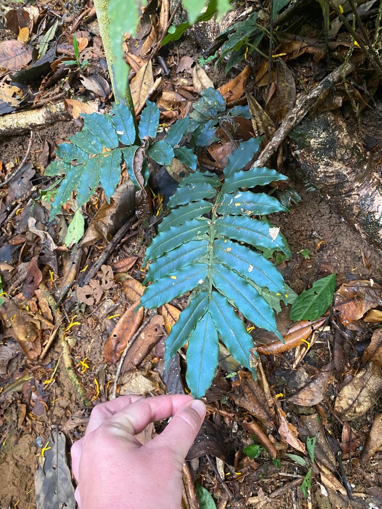
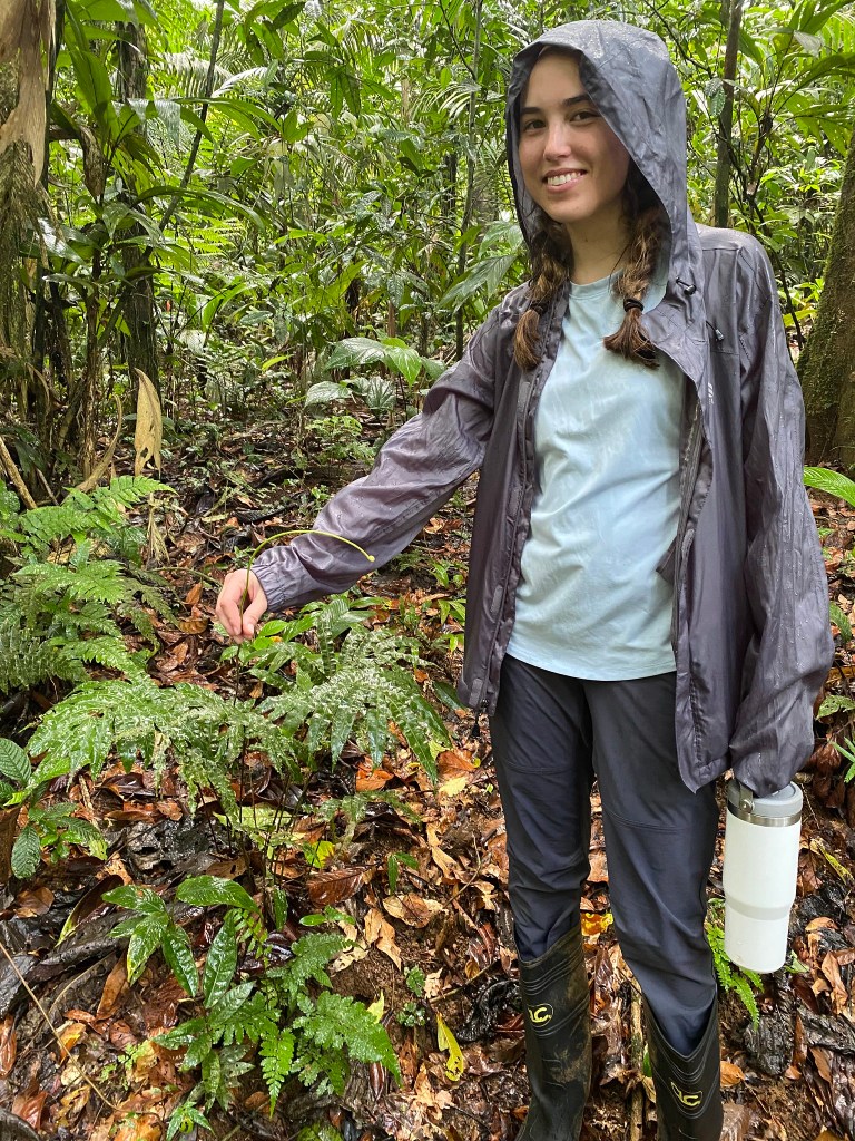
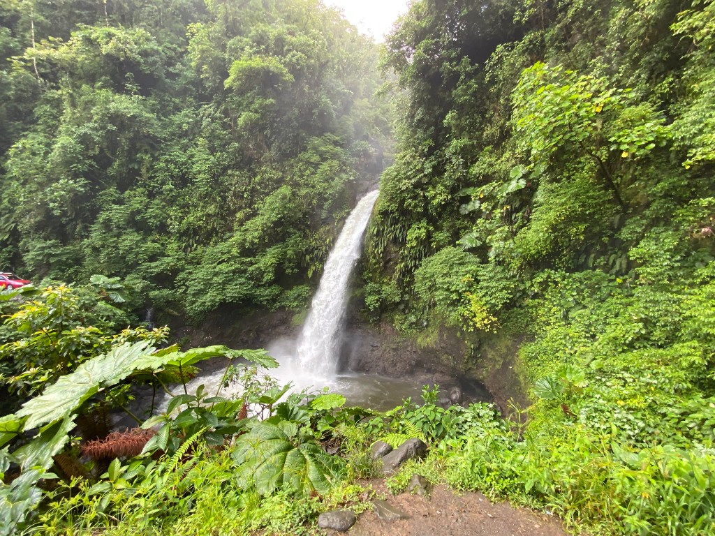
Last May, my student Jordyn Regier and I went to Costa Rica to investigate opportunities for future research projects. We found and identified so many ferns! We spent about a week hiking through the jungle and collecting preliminary data. I can’t wait to return!
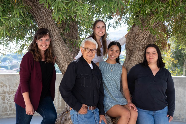
Drs. Helen Holmlund and Stephen Davis with 2021 SURB students: Mayra Hernandez, Jordyn Regier, and Camille Sicangco
A couple weeks ago, three students in my lab (Mayra Hernandez, Jordyn Regier, and Camille Sicangco) presented their research at the Southern California Conference for Undergraduate Research (program available here). They presented the projects that they initiated last summer during Pepperdine’s SURB program (our NSF REU Site Program).
Mayra presented her research on coastal sage scrub and vegetation type conversion in the chaparral. Jordyn presented her project on chaparral fern gametophyte desiccation tolerance. Camille presented her research on how vegetation type conversion affects soil respiration in the chaparral. Stay tuned as these folks continue their research!
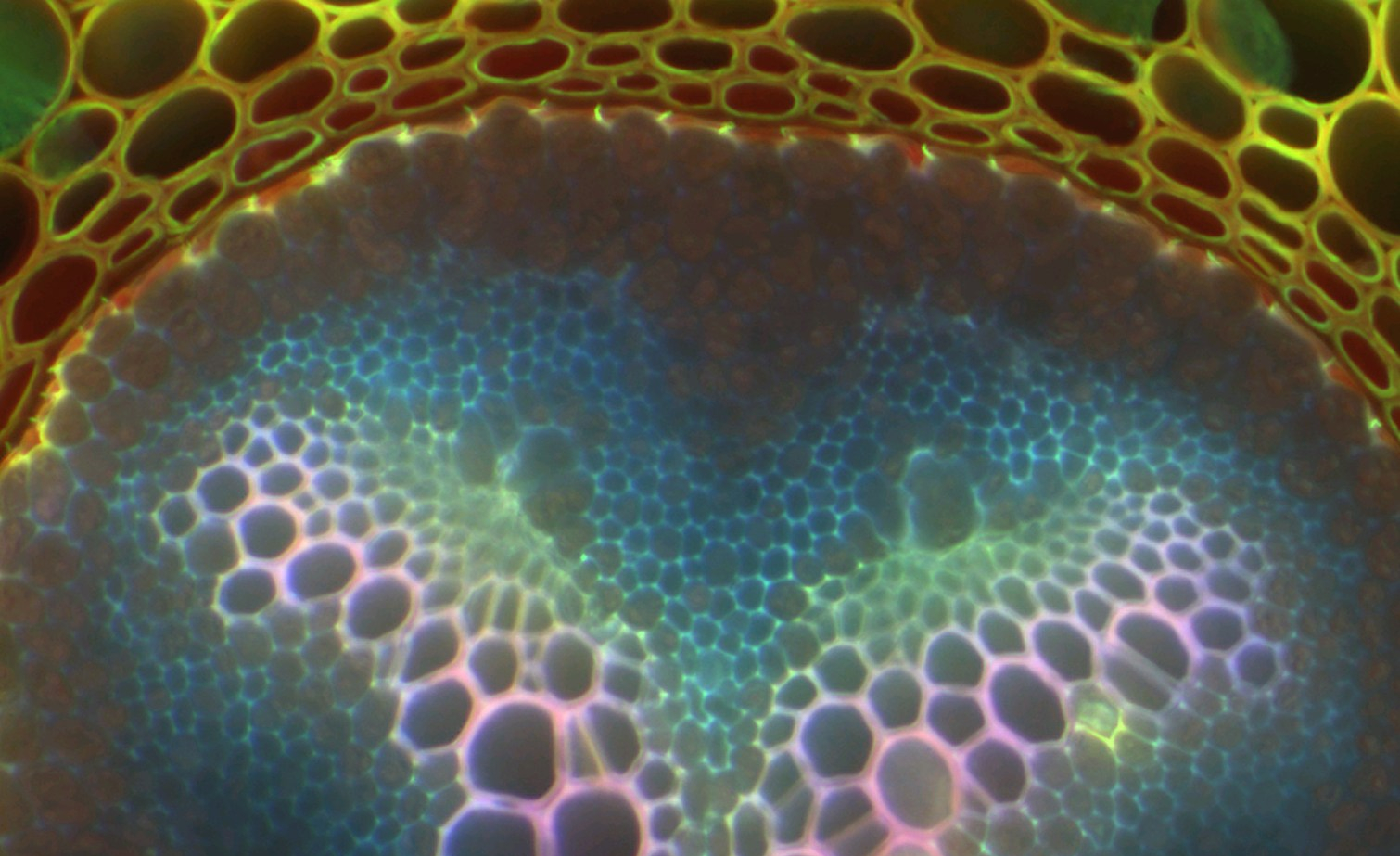
New Phytologist’s blog recently featured our 2019 research article on the resurrection ferns. The blog post features a fluorescence micrograph of a resurrection fern stem by Dr. Anna Jacobsen (shown above). Click on the link below to read the blog post.
Holmlund, H.I., Pratt, R.B., Jacobsen, A.L., Davis, S.D. and Pittermann, J., 2019. High‐resolution computed tomography reveals dynamics of desiccation and rehydration in fern petioles of a desiccation‐tolerant fern. New Phytologist 224(1): 97-105.
Last summer (winter?) in Australia, I traveled to Lizard Island to study the ferns there. Lizard Island is located off the coast of northeastern Australia in the Great Barrier Reef.
Lizard Island seen from the plane
Flying over the Great Barrier Reef
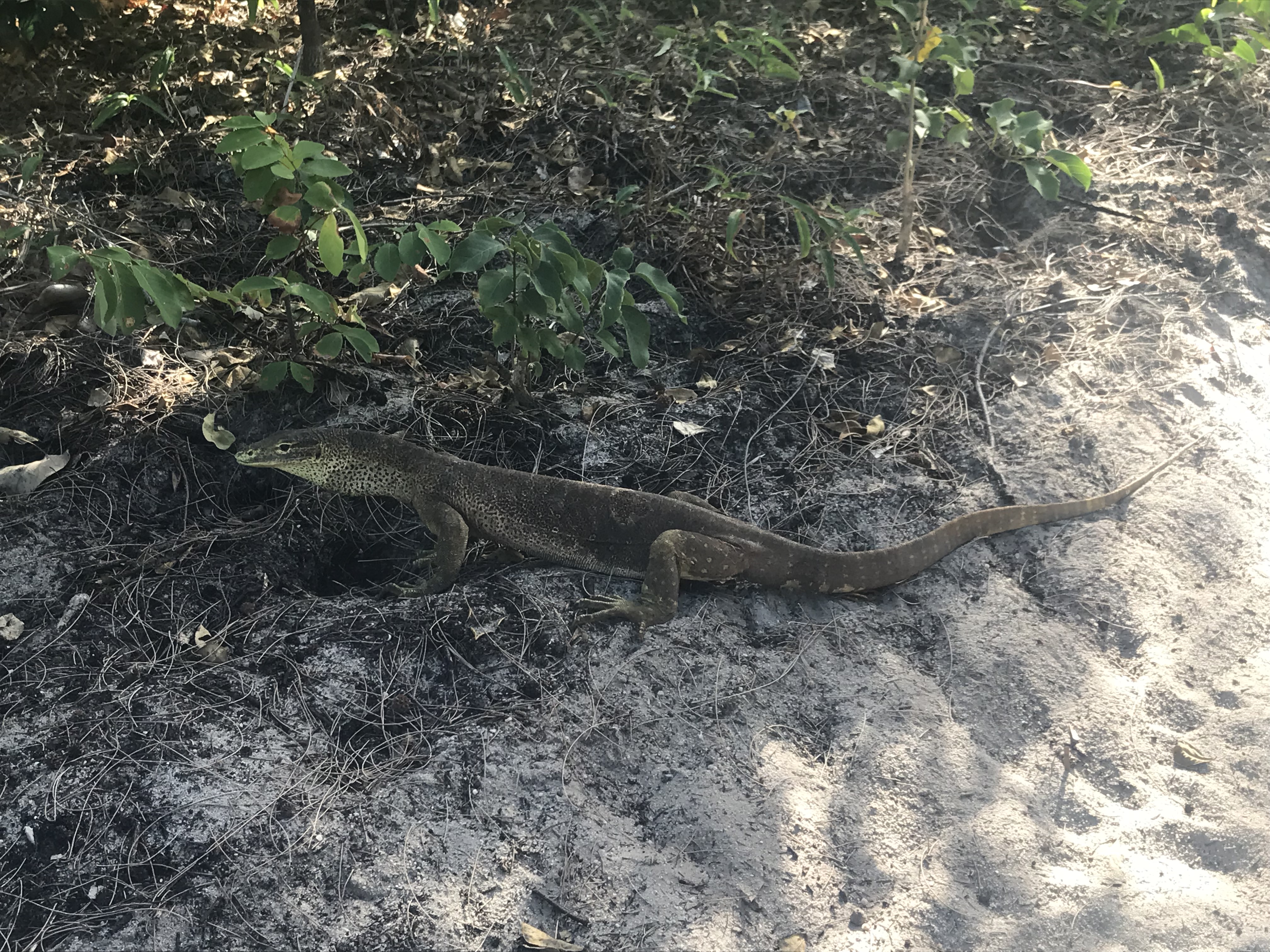
Lizard Island was named by Captain Cook because of the large goannas that roam the island.
As far as field work goes, it was a pretty comfortable place to work.
Loomis Beach
View from my cabin
There is a small swamp in the middle of the island, and that is where my ferns are located. The only way around the island is on foot, so I carried my gear with me.
Trail along the swamp
Carrying the LI-6400 to my site
My gear
Measuring photosynthesis
The mangrove ferns grow along the edges of the swamp, among the mangrove tree roots. They are difficult to spot and were only recently discovered on Lizard Island.
Mangrove ferns up close
Mangrove ferns between the swamp and the Pandanus
While measuring water salinity, I had to be mindful of the crocodiles. Various precautions were necessary…
Sampling salinity at a distance
Sampling from the bridge
The bridge/boardwalk through the swamp
The community of students and researchers on Lizard Island makes it a lovely place to work! Saturday beach barbecue is a time-honored tradition.
Even in my lab, I was never alone.
World’s tiniest praying mantis crawling along my phone cord
“Harry” the huntsman
My measurements were successful, and I’m analyzing the data now. Stay tuned for what I find!
This past year, I received a NSF GROW fellowship to study ferns in Marilyn Ball’s lab at the Australian National University. I was based in Canberra, the “bush capital,” for most of my time in Australia.
ANU is indeed situated in “the bush”! The university is surrounded by hills covered with native plants and wildlife.
View from the trail on Black Mountain
Echidna spotted on Black Mountain
Brindabella Mountains
Canberra is a great place for bird-watching! The cockatoos and parrots are everywhere.
sulfur-crested cockatoo in Canberra, Australia
sulfur-crested cockatoos
corellas
gang-gang cockatoo
galahs
King Parrot
King Parrots on ANU campus
I really enjoyed my time in Canberra. Stay tuned for the findings from my project!
Our study of how water moves through resurrection ferns was recently published in New Phytologist! This study was a collaboration with Drs. Anna Jacobsen and Brandon Pratt at California State University, Bakersfield.
We used high resolution micro-CT scans to visualize the dynamics of water movement in resurrection ferns while they were drying out and resurrecting. We combined this approach with light and fluorescence microscopy to learn which anatomical traits may facilitate desiccation tolerance in these ferns.
The abstract is available here, and requests for full text can be sent to helenireneholmlund at gmail dot com.
Dr. Jacobsen and Dr. Pratt’s websites are available here and here.
Desiccated Pentagramma triangularis next to a 3D microCT reconstruction of its stipe. PC: Dr. Anna Jacobsen
Pentagramma triangularis in the chaparral understory. The shorter leaves (curling) are starting to dry out. PC: Dr. Steve Davis
Our lab recently had a short paper (a disease “note”) accepted to the journal Plant Disease! The paper is titled “First Report of Botryosphaeria dothidea Causing Stem Canker and Plant Death in Malosma laurina in Southern California.” Pepperdine graduate (class of 2017) Natalie Aguirre is the lead author on the paper.
Natalie examining an infected Malosma laurina
Over the last couple years, Natalie has investigated a serious fungal infection in Malosma laurina, a co-dominant chaparral shrub species on Pepperdine campus. M. laurina (laurel sumac) is an important member of the chaparral plant community throughout the Santa Monica Mountains in Malibu, CA. In the past, this shrub has been very resistant to the effects of drought because of its deep roots that access deep water resources. While shallow-rooted plants experience dehydration, M. laurina has remained relatively hydrated.
Natalie standing inside a healthy M. laurina
However, in 2015 we noticed severe dieback in M. laurina along the coastal exposures of the Santa Monica Mountains. A fungus appears to be blocking water transport in the vascular system, cutting off water supply to the leaves. We suspect that the extended drought of 2012-present may have pre-disposed the plants to be more susceptible to this infection, perhaps by weakening their immune systems or limiting their carbon resources. In the photo below, you can clearly see where the point in the stem where the fungus is growing and blocking water supply to the leaves.
Measuring photosynthesis in infected M. laurina
B. dothidea growing in a Petri dish
Check out Natalie’s paper when it becomes available online! Also, she has presented this research at the Ecological Society of America (2017) and the Botanical Society of America (2016). You can read her abstracts here:
There are many student authors on this paper because this was a collaboration of many people in Dr. Davis’ lab! His Plant Physiology class (fall 2015) conducted their class projects on the M. laurina infection. Thanks to everyone’s hard work, we learned a lot about the nature of the fungal infection.
Plant Physiology class excavating M. laurina roots
Plant Physiology class poster session
Natalie is currently working on a research Fulbright in Madrid, Spain. We are excited to see the great things in store for her!
Earlier this month, several of us took a field trip to Santa Cruz Island to continue our research project on foliar water uptake in island ferns. This time, we were transplanting island ferns to the mainland for further experiments.
As always, the boat ride was fantastic! Dolphins followed us part of the way, and the fog was clearly visible around the island as we approached.
Anacapa Island
Prisoners’ Harbor
The island has many different microsites with small differences in climate. Thus, we needed to travel all over the island to locate our eight diverse fern species!
Driving around the island
Leaving the field station
Many of the fern species were located near the west end of the island in Christy Pines. This seems like a logical place for the ferns to grow because this west end of the island is frequently foggy. After collecting the ferns, we continued past Christy Pines to Christy Beach.
Gaby at Christy Beach
The next day, we drove back to the dock at Prisoners’ Harbor. From there we hiked to Pelican Bay, where the rest of our fern species grow. There is a small perennial water source near Pelican Bay, and many of the dehydration sensitive evergreen ferns grow there.
The lookout house above Prisoners’ Harbor
Pelican Bay
Looking back at Prisoners’
Cristian beside the century plants at Pelican Bay
It was certainly a successful trip! I am excited for the experiments we will run over the next few months, and I am definitely looking forward to returning to the island.
Last August, I went to Oregon with my labmate Kate to view the solar eclipse! The journey was a fantastic adventure, and we enjoyed every minute of it.
First, we drove through Eureka and Crescent City in northern California. The air was filled with smoke from the nearby wildfires, creating a fiery sunset. Also, we drove past an enormous herd of elk.
We camped in the redwood forest, alongside the ferns and banana slugs.
Redwoods and ferns in Eureka, CA
Next, we drove by Crater Lake, which was also hazy from all the smoke, but still incredibly blue!
We camped that night at a place called Cinder Hill campground.
We were worried about traffic on the Oregon highways, so we left at 3 am! However, we got up extra early to make coffee.
Actually, there was no traffic on the way into Madras area to view the eclipse. We decided to view the eclipse from Cove Palisades campground and waited there. Cove Palisades alone was worth the trip! The state park is located at the junction of the Deschutes and Crooked rivers.
We were joined by many fellow eclipse chasers. The eclipse itself was breath-taking, like nothing I had ever seen!
Driving back to Eureka was when we hit the traffic. Our trip was extended from 7 hours to about 13 hours!
We stayed at Patrick’s Point State Park after that. The park has coastal redwoods growing near a beach.
Rolling up a wet tent is hard!
Overall, the trip was fantastic. I hope I can go see the next eclipse!
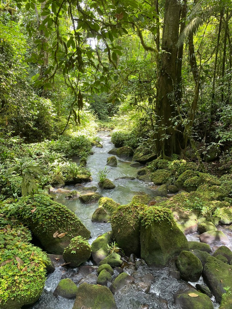
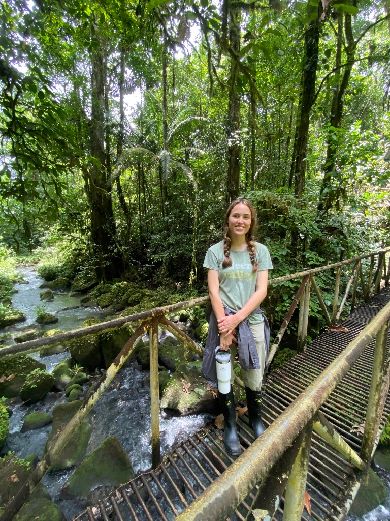
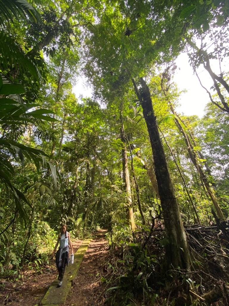
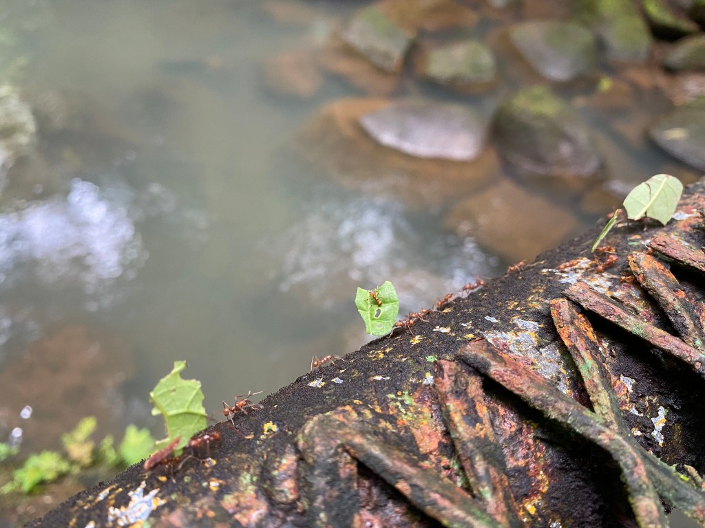
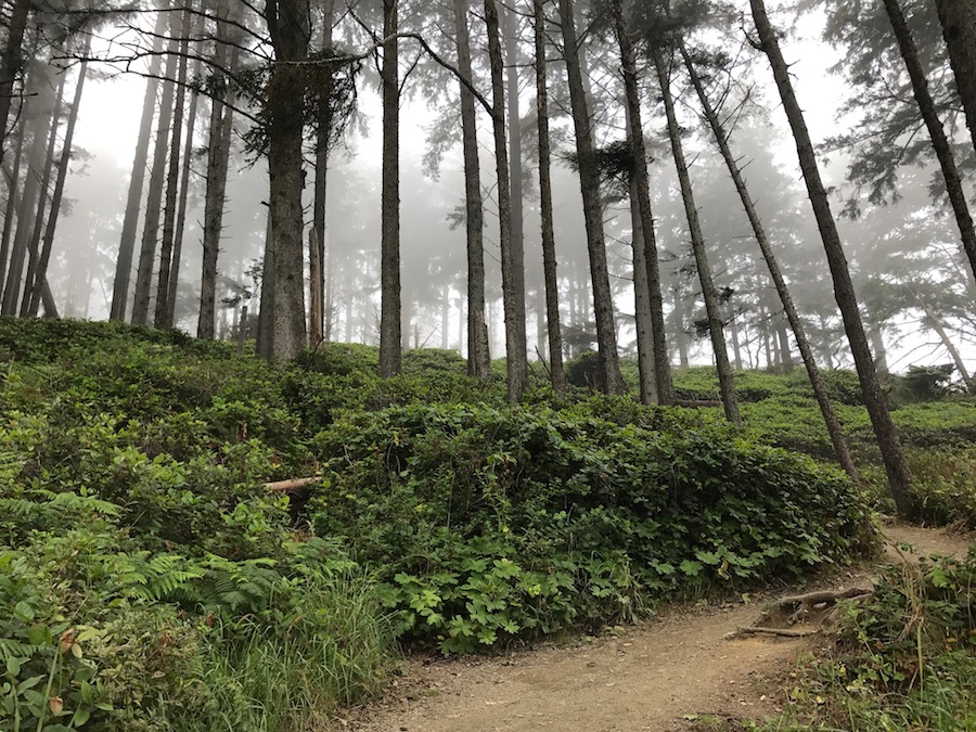
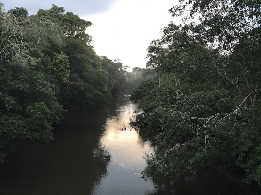
This summer, I spent three weeks doing field work at La Selva Biological Research Station in Costa Rica!
The field station is located in the beautiful Costa Rican rainforest.
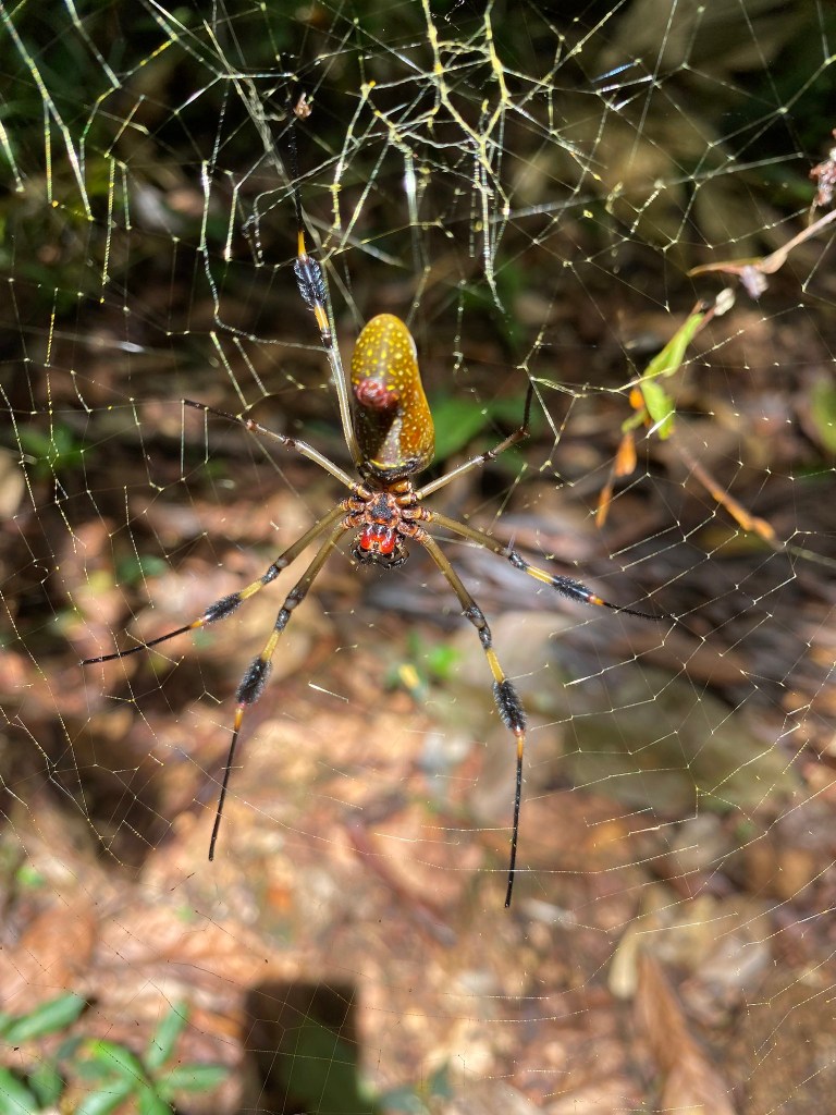
The rainforest is alive with frogs, bugs, and many other critters!
scarlet-webbed tree frog, Costa Rica
The howler monkeys woke us up every morning.
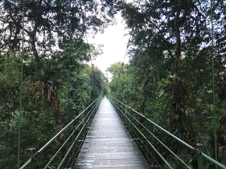
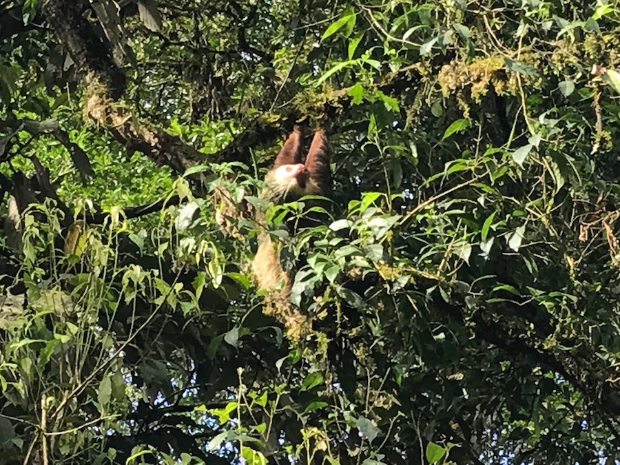
Sloths are hard to spot in the trees, but this one liked to hang out near the bridge.
Deadly eyelash vipers live in the forest.
Eyelash viper
Of course, the plants were the most exciting part of the rainforest.
I’m climbing a tree!
At the top of the viewing tower at La Selva in Costa Rica
Not a plant, but still cool
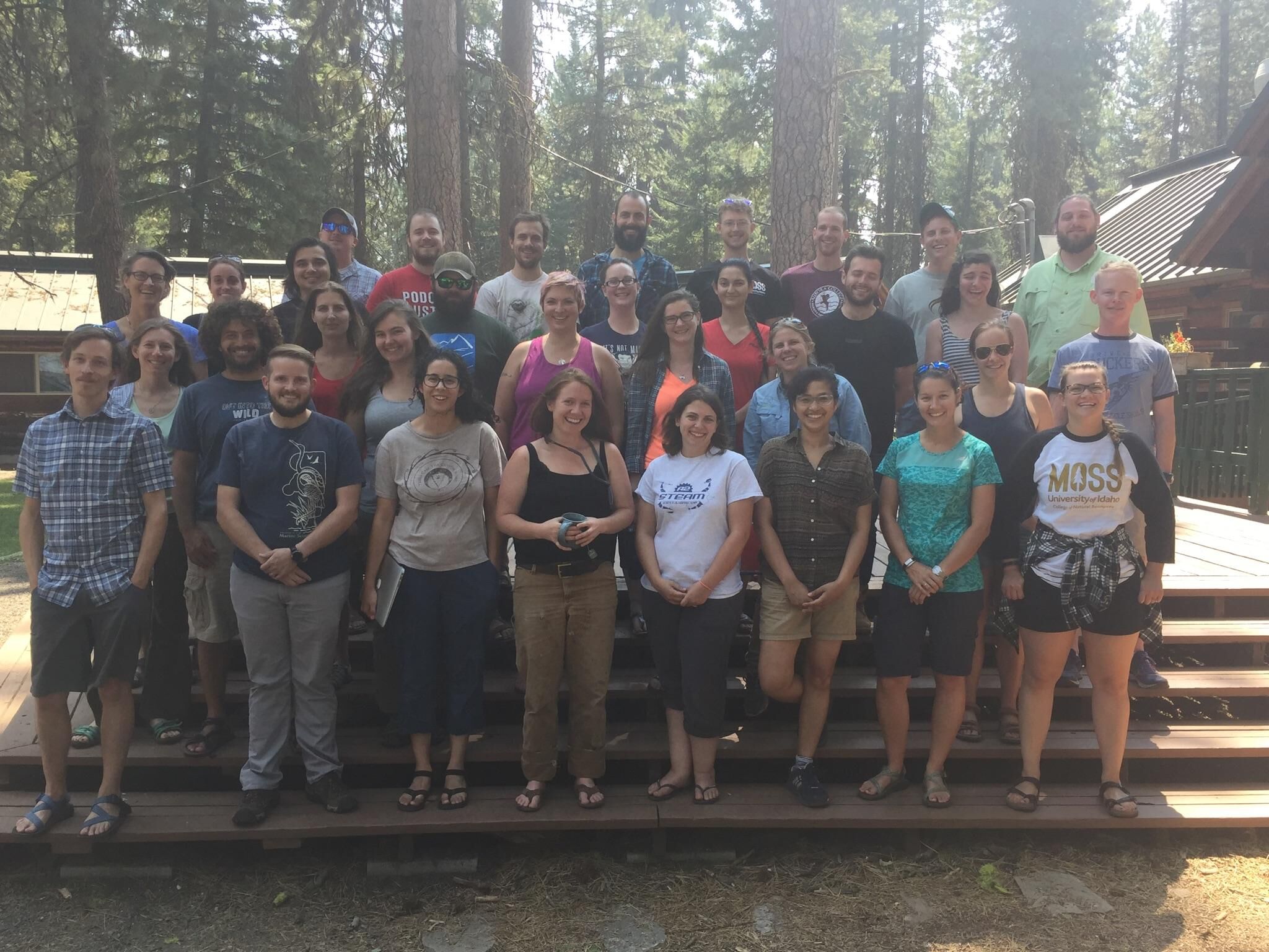
In August, I participated in a Plant Hydraulics methods workshop in McCall, Idaho. We met for several days at the MOSS field station and learned various techniques related to plant hydraulics.
I flew straight from Idaho to Portland, OR, to present my research at the Ecological Society of America.